

The optical impression is an excellent alternative to impressions taken with the use of special impression materials. In our dental clinic we have been using this method since 2010 when performing all works related to CAD / CAM technology: fully ceramic bridges, crowns, inlays, onlays and crowns on implants. In the article below, we will describe the types of impressions used in dentistry.
Prosthetic impressions
Taking dental impressions has long been associated with all dental reconstruction work.
What are dental impressions for?
Impressions are used to accurately “copy” the surface of the mucosa and teeth. On the basis of the impression, a plaster model is made, which in detail reproduces the shape of the teeth, alveolar appendages and the mucosa.
Using the model, you can design and produce prosthetic restorations, such as crowns, bridges, dentures and other.
Classic impressions
For many years different impression materials and ways of taking impressions have been used. Classic prosthetic impressions have not lost their importance. Technology of production of impression materials dedicated to various types of prosthetic work is constantly evolving. However, taking a good impression is always associated with discomfort on the part of the patient and is often impossible due to a strong gag reflex. The doctor taking the impression also needs to demonstrate a high level of skill in order to accurately reproduce the shape of the oral cavity.
Optical impressions
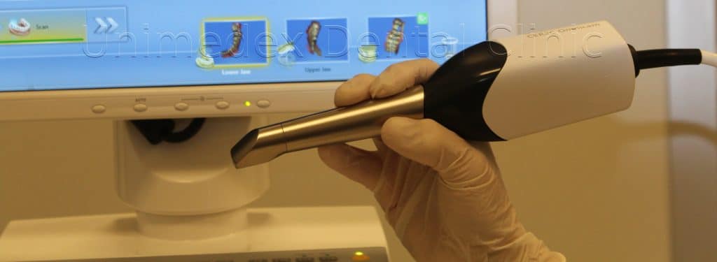
An optical impression is a common name.
The correct technical name is a multidimensional scan of the oral cavity.
The basis for the application and development of CAD / CAM technology for dentistry was the design and introduction of an intraoral scanner.
The technology was developed at the University of Zurich and its pioneer was Sirona Gmbh (now Dentsplay Sirona), which in 1980 introduced the CEREC (“Chairside Economical Restoration of Esthetic Ceramics”) technology.
CEREC Intraoral Scanner (link) is a tool with which the doctor takes many pictures of the dentition in a short span of time, which are then put together into one three-dimensional spatial image on a computer. The dentist can see all the important details of the optical impression in a high magnification on a screen and, if necessary, improve the “preparation” of the teeth for the crown fitting. The physical prosthetic model hitherto cast by the technician is unnecessary, because a model generated by a computer program is sufficient. Based on the digital model, many prosthetic reconstructions can be designed both on-site, in the dentist’s office, as well as in a remote dental laboratory, where the model can be sent via e-mail.
All this saves a huge amount of time! It became possible to make a crown or a bridge in just one visit!